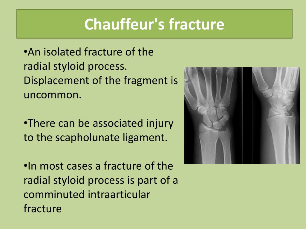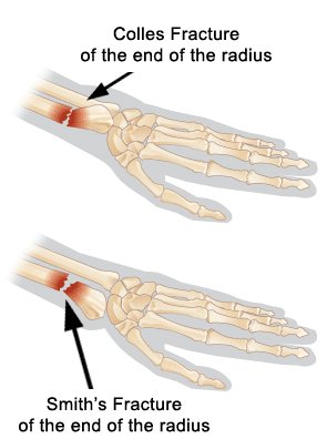

In general, radiographic interpretation should comment on the presence of a distal radial fracture with volar angulation, the fracture location (extra-, juxta-, or intra-articular), the degree of angulation, and the degree of displacement. In these scenarios, a CT can help define, and best appreciate not only the pattern of injury but also help the surgeon to plan for operative reduction strategy. In certain instances, a CT may be useful in situations of extensive comminution or intra-articular fracture patterns. The latter is rarely necessary in most clinical situations. However, additional radiographs of the wrist such as traction, oblique, and fossa lateral views along with advanced imaging, may offer vital information about the injury to include associated soft tissue injuries. Typically, orthogonal views (AP, lateral) are adequate.
Barton vs colles fracture series#
Ī wrist radiograph series is adequate for the characterization of a distal wrist fracture and can differentiate between a Colles and Smith fracture. Another cause of neurovascular compromise seen in Smith fractures and other distal radius fractures is acute compartment syndrome of the forearm. Less commonly, but still present neurological concerns include both radial and ulnar nerve compression. Up to 15% of these fractures may show symptoms of acute carpal tunnel syndrome (ACTS) from compression to the median nerve. A compromise would necessitate immediate attempts at closed reduction. Įvaluation of the extremity's neurovascular status is imperative. There may also be an association of ulnar styloid base fractures. In addition to the volar displacement of the distal fragment, disruption of the distal radioulnar joint (DRUJ) and the triangular fibrocartilage complex (TFCC) often occurs.

One of Smith’s first diagnostic criteria was a deformed wrist with swelling visible on the volar side and the prominence of the ulna along the dorsum of the wrist. Also, present on the exam are swelling, pain, and decreased ROM.

The physical exam may reveal a deformity of the distal forearm, but the direction of angulation- dorsal (Colles) or volar (Smith) is difficult to discern on visualization. Data appears to support a direct correlation between low-energy trauma-induced distal radius fracture and decreased bone mineral density. Between the ages of 64 to 94, women are six times more likely than men to sustain this type of fracture. In the elderly population, distal radial fractures are the second most common fracture, second only to hip fractures. Almost all distal radius fractures arise in children sustaining high-energy falls and osteoporotic seniors who suffer low-energy falls. The highest incidence of Smith's fractures is in young males and elderly females. However, Smith fractures make up approximately 5% of all radial and ulnar fractures combined.

Distal radial fractures are the second most common fracture in the elderly. With over 600000 cases annually in the United States alone, distal radial fractures account for more than 16% of all adult fractures and 75% of forearm fractures. The distal radius is the most common fracture site in the upper extremity.


 0 kommentar(er)
